مقارنة بين تقنية PCR وتقنية qPCR في تشخيص أمراض الدواجن
مقالة عن تقنیات المولکیولر بایولوجي الحدیث التطبیقي

للدکتور ماجد حمید الصایغ / استرالیا
8/ 9/ 2025
مقدمة
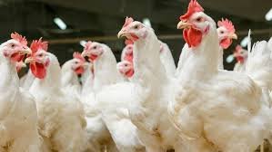
تُعد التقنيات الجزيئية مثل تفاعل البوليميراز المتسلسل (PCR) حجر الزاوية في تشخيص العديد من أمراض الدواجن الفيروسية والبكتيرية. ومع تطور التكنولوجيا، ظهر نوع متقدم من هذه التقنية يُعرف باسم التفاعل المتسلسل الكمي في الوقت الحقيقي (qPCR) الذي يتيح الكشف عن المادة الوراثية بشكل لحظي وكمي خلال عملية التضخيم. لقد أصبح كل من PCR التقليدي وqPCR أدوات أساسية في مختبرات التشخيص البيطري لتحديد مسببات الأمراض بسرعة ودقة (mdpi.com) ، مما يساعد في السيطرة على أمراض خطيرة مثل الإنفلونزا الطيرية (AI) ومرض نيوكاسل (NDV) والتهاب كيس فابريشيا (IBDV) وفيروسات الأدينو في الدواجن (FAdV) والمايكوبلازما جاليسبتيكوم/سينوفي (MG/MS). نستعرض في هذا التقرير الشامل الفروقات بين PCR التقليدي وqPCR من حيث الحساسية والنوعية والوقت والحاجة إلى التسلسل الجيني، واستخدام كل منهما مع أنواع العينات المختلفة، وتطبيقاتهما عبر الأمراض المذكورة، إضافةً إلى دقة النتائج (بما في ذلك الضوابط الداخلية) وأهم التوصيات العملية للمختبر والميدان.
Comparison between Conventional PCR and Real-Time PCR (qPCR)
Conventional PCR differs from qPCR in several key aspects related to diagnostic performance and requirements:
First: Sensitivity: qPCR is more sensitive than conventional PCR. The real-time monitoring of amplification in qPCR allows the detection of very small amounts of the target DNA/RNA, which conventional PCR may fail to detect (phchd.com). In other words, qPCR can detect very few copies of the target gene, thanks to its ability to track the accumulation of the product during the early stages of the reaction. This is particularly useful for diagnosing samples with low levels of the pathogen (such as early stages of infection or diluted environmental samples).
Second: Specificity: In conventional PCR, the identification of the amplification product is often based on the size of the DNA fragment visualized on an agarose gel, which can sometimes allow for non-specific amplification. In contrast, qPCR uses specific fluorescent probes or dyes that bind only to the target amplification product, thus increasing the specificity of the reaction (phchd.com). For example, TaqMan probes in qPCR provide an additional layer of verification (as they must match the internal sequence of the product), so a signal is only detected when the correct target is amplified. This significantly reduces false-positive results compared to conventional PCR or even qPCR using general dyes like SYBR (Greenmdpi.com). Therefore, qPCR provides a higher degree of certainty that the detected signal corresponds to the specific pathogen of interest. Third: Speed and time: Conventional PCR typically takes several hours to complete amplification and requires additional steps after the reaction to detect the results (such as electrophoresis and gel analysis to visualize the DNA fragment). These additional steps increase the time required to obtain results and increase the risk of contamination. qPCR, on the other hand, provides faster results by detecting the product during amplification, without the need for any post-reaction steps; the result is determined as soon as the reaction is complete (phchd.com). In many cases, results can be obtained within one to two hours using qPCR, compared to ~3-4 hours for conventional PCR, including preparation, amplification, and gel electrophoresis. Furthermore, qPCR operates in a closed system (a tube that is not opened after the reaction), reducing the risk of contamination and saving time that might be spent on retesting due to sample contamination (phchd.com).
Fourth: The need for sequencing: In conventional PCR, the amplification product is a DNA fragment visualized on a gel; to definitively identify this product (especially when diagnosing viruses or bacteria with multiple strains), sequencing of the amplified product or analysis using techniques such as Restriction Fragment Length Polymorphism (RFLP) may be necessary (pmc.ncbi.nlm.nih.gov). This confirmatory sequencing precisely determines the nucleotide sequence of the product to verify its match with the target pathogen and to compare the isolate with known strains. For example, conventional PCR was combined with sequencing in the diagnosis of avian adenovirus to identify different serotypes due to the high genetic diversity of these viruses (mdpi.com). In contrast, qPCR typically relies on specific probes designed to target a particular region of the pathogen’s genome. If the probes and primers are carefully designed, a positive qPCR result provides strong confirmation of the presence of the target sequence, often obviating the need for immediate gene sequencing. However, gene sequencing remains essential in certain situations, such as confirming the identity of an unusual strain, identifying important genetic traits (e.g., genes associated with virulence or resistance), or for epidemiological and research purposes following initial diagnosis. In other words, qPCR offers rapid and specific diagnosis, but it may not replace sequencing when a more detailed characterization of the detected strain is required (e.g., for genotyping or monitoring the emergence of new mutations). (mdpi.com).
Using the Two Techniques with Different Sample Types
The success of PCR or qPCR detection depends on the type of sample collected and the method used for collection. Below is an explanation of how PCR and qPCR are used with different types of poultry samples:
1. Tissue Samples: Both conventional PCR and qPCR can be applied to infected tissue samples (such as internal organs) to detect the pathogen. This requires extracting DNA (for DNA viruses and bacteria) or RNA (for RNA viruses, with a reverse transcription step) from the tissue. In some diseases, tissue is the preferred sample: for example, Bursa of Fabricius tissue is preferred when suspecting Infectious Bursal Disease (IBD), and liver tissue is preferred for diagnosing Feline Adenovirus (FAdV) infection. Both techniques can detect the presence of the pathogen’s genetic material in the tissue, but qPCR is more sensitive, especially when low levels of the virus are present in the tissue sample. qPCR also allows for quantification of the viral load in the tissue, which can help assess the severity of the infection. However, tissue extracts may contain inhibitors of the reaction, so it is important to use internal controls, as discussed below, to ensure accurate results with both PCR and qPCR.
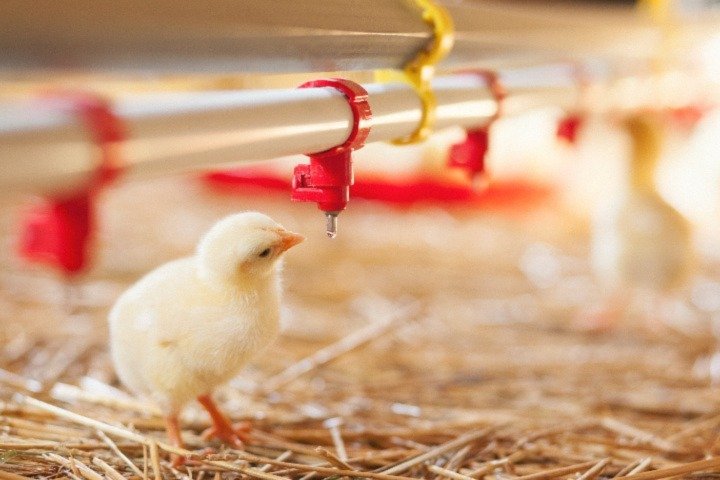
2. Swabs: Swabs are a fundamental sampling tool, especially for respiratory and enteric diseases. Examples of swabs include tracheal or oropharyngeal/laryngeal swabs (for sampling the upper respiratory tract) and cloacal swabs (for sampling the gastrointestinal tract or detecting viral shedding in feces). Both PCR and qPCR are used on the extracted DNA/RNA from the swabs to detect the presence of the pathogen. Since the amount of pathogen in swab samples may be limited, qPCR is often preferred for swab testing due to its higher sensitivity and ability to detect even low viral or bacterial loads (phchd.com). Studies have shown that real-time PCR significantly increased the detection rate of certain viruses in swab samples compared to conventional PCR on the same samples (sciencedirect.com). For example, in the diagnosis of Newcastle disease in poultry, qPCR showed a higher positive detection rate than conventional PCR, confirming its superior diagnostic accuracy (sciencedirect.com). qPCR also excels in its ability to process large numbers of samples more quickly (using 96-well plates, for example), making it suitable for large-scale farm surveys or rapid epidemiological surveillance programs. In general, swabs (especially respiratory swabs) are the primary samples for testing diseases such as AI, NDV, and MG/MS, and it is always recommended to collect them correctly (taking a deep, gentle swab) and place them in a suitable transport medium to maintain sample integrity until they reach the laboratory.
3- Environmental samples: Molecular techniques can be used to detect pathogens in environmental samples collected from the air or surfaces of poultry houses. Accumulated dust in poultry housing is a prime example of environmental samples used in diagnostics; samples of dust or shed feathers are collected and tested by PCR for the genetic material of viruses or bacteria circulating in the flock. PCR tests using dust samples are advantageous because they are easy to collect (farm workers can collect them without disturbing the birds) and are non-invasive. They also provide a good representation of the overall health status of the flock, rather than requiring samples from each individual bird (journals.plos.org). Dust sampling using qPCR has been used to monitor the spread of vaccines or specific viruses (such as monitoring the efficacy of the Infectious Laryngotracheitis (ILT) vaccine, or detecting the presence of viruses like IBV and NDV) and has shown promising results (journals.plos.org). Another advantage of dust samples is that viral genetic material remains stable and detectable for extended periods; studies have shown that the genomes of viruses like IBV can remain detectable in stored dust for up to four months at 37°C (journals.plos.org). However, it should be noted that the virus itself may no longer be infectious after this time, even though its genetic material remains detectable. In general, qPCR allows for highly sensitive detection in environmental samples with very low DNA concentrations, increasing the chances of detecting even small amounts of pathogens in the barn environment or ventilation systems. This method is promising for routine disease monitoring in large poultry farms as part of biosecurity programs, and should be used in conjunction with direct samples from the birds for further confirmation.